In the ever – evolving landscape of medical technology, glucose biosensors stand as a remarkable testament to the fusion of biology, chemistry, and electronics. These compact yet sophisticated devices have revolutionized the way we monitor blood glucose levels, providing individuals with diabetes and healthcare professionals with a rapid, accurate, and convenient means of assessment. From the moment a tiny drop of blood makes contact with the sensor to the instant a glucose reading is displayed, a series of highly coordinated processes occur, leveraging the principles of biochemistry and electrochemistry.
The Fundamental Components of Glucose Biosensors
Sensing Element: The Heart of the Biosensor
At the core of every glucose biosensor lies the sensing element, which is responsible for detecting glucose molecules in the blood sample. The most commonly used sensing element is an enzyme called glucose oxidase or glucose dehydrogenase. Glucose oxidase catalyzes the oxidation of glucose in the presence of oxygen, producing gluconic acid and hydrogen peroxide. Glucose dehydrogenase, on the other hand, uses a co – factor to oxidize glucose, resulting in a different set of reaction products. These enzymes are highly specific to glucose, meaning they selectively react with glucose molecules while ignoring other substances in the blood, ensuring the accuracy of the glucose measurement.
The enzymes are immobilized on a suitable substrate, such as a porous membrane or a conducting electrode surface. This immobilization process is crucial as it allows the enzymes to remain in place and interact with the glucose molecules in the blood sample without being washed away or degraded. The choice of substrate and the method of enzyme immobilization can significantly impact the performance and lifespan of the sensing element.
Transducer: Converting Biological Signals into Electrical Signals
Once the glucose molecules have reacted with the enzyme on the sensing element, the resulting chemical change needs to be converted into an electrical signal that can be measured and processed. This is the role of the transducer. There are several types of transducers used in glucose biosensors, with electrochemical transducers being the most prevalent.
In electrochemical transducers, the chemical reaction involving glucose oxidation generates an electrical current or a change in voltage. For example, in amperometric transducers, the production of hydrogen peroxide during the glucose oxidase – catalyzed reaction can be detected electrochemically. When an appropriate voltage is applied to the electrode, the hydrogen peroxide is oxidized, generating an electrical current. The magnitude of this current is directly proportional to the concentration of glucose in the blood sample, allowing for the quantification of glucose levels.
Potentiometric transducers, on the other hand, measure the change in electrical potential (voltage) that occurs as a result of the glucose – enzyme reaction. The change in potential is related to the concentration of reaction products or the consumption of reactants, which can be correlated with the glucose concentration.
Signal Processor: Analyzing and Interpreting the Data
The electrical signal generated by the transducer is often weak and noisy, and it requires further processing to obtain an accurate glucose reading. This is where the signal processor comes into play. The signal processor typically consists of an amplifier, which boosts the strength of the electrical signal, and a filter, which removes unwanted noise and interference.
Once the signal is amplified and filtered, it is sent to an analog – to – digital converter (ADC). The ADC converts the continuous analog electrical signal into a discrete digital signal, which can be easily processed by a microprocessor. The microprocessor uses a pre – programmed algorithm to analyze the digital signal and calculate the glucose concentration based on the relationship between the electrical signal and the known glucose concentration – signal response curve. This curve is established during the calibration process of the biosensor.
The Step – by – Step Process of Glucose Detection
Sample Application
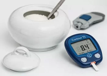
The process of glucose detection begins with the application of a blood sample to the glucose biosensor. In most cases, a small drop of blood, usually obtained by pricking the finger with a lancet, is applied to the sample application area of the test strip. Test strips are an integral part of many glucose biosensors, as they contain the sensing element and are designed to facilitate the interaction between the blood sample and the sensor.
Capillary action often plays a crucial role in drawing the blood sample into the test strip. The narrow channels and pores within the test strip are designed to draw the blood in, ensuring that the sample comes into contact with the sensing element evenly and efficiently.
Enzymatic Reaction
Once the blood sample is inside the test strip, the glucose molecules in the blood immediately start to react with the immobilized enzyme on the sensing element. As mentioned earlier, if glucose oxidase is used, glucose is oxidized to gluconic acid and hydrogen peroxide. The rate of this reaction depends on the concentration of glucose in the blood sample; the higher the glucose concentration, the faster the reaction proceeds, and the more reaction products are generated.
Signal Generation and Detection
As the enzymatic reaction takes place, the resulting chemical changes are detected by the transducer. In electrochemical biosensors, the production of reaction products such as hydrogen peroxide leads to the generation of an electrical current or a change in voltage at the electrode surface. The transducer measures this electrical signal and converts it into a form that can be further processed.
Data Processing and Display
After the electrical signal is amplified and filtered, it is converted into a digital signal by the ADC and sent to the microprocessor for further analysis. The microprocessor uses the pre – established calibration curve to convert the electrical signal into a glucose concentration value.
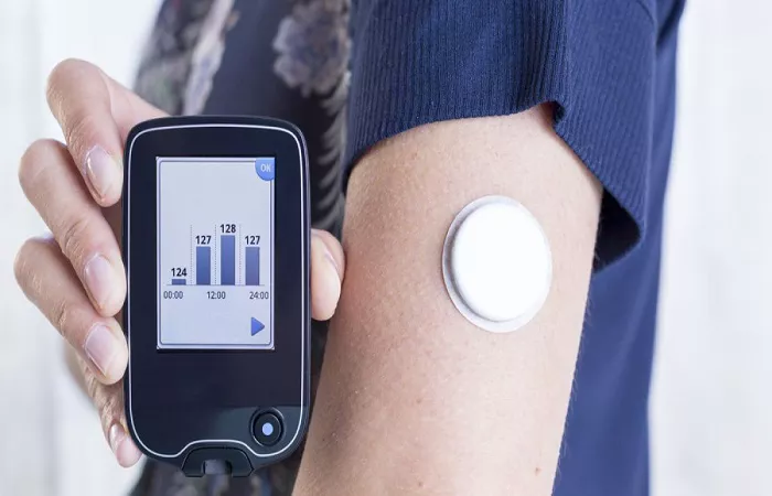
Once the glucose concentration is calculated, it is displayed on the screen of the glucose biosensor device. The display is typically clear and easy to read, showing the glucose level in milligrams per deciliter (mg/dL) or millimoles per liter (mmol/L), depending on the region and the settings of the device. Some advanced glucose biosensors may also store the glucose readings, allowing users to track their glucose levels over time and generate reports for themselves or their healthcare providers.
Calibration and Quality Control of Glucose Biosensors
Calibration: Ensuring Accuracy
Calibration is a critical step in the operation of glucose biosensors. Since the relationship between the electrical signal generated by the transducer and the actual glucose concentration can vary due to factors such as differences in enzyme activity, electrode performance, and manufacturing variations, calibration is necessary to establish a reliable correlation.
Most glucose biosensors come with calibration codes, which are unique numbers that correspond to the specific batch of test strips being used. These calibration codes are entered into the biosensor device, and the device uses them to adjust its internal settings and algorithms to account for any differences in the performance of the test strips. Additionally, some biosensors may require calibration using known glucose standards, which are solutions with precisely defined glucose concentrations. By comparing the measured values of these standards with the known values, the biosensor can be calibrated to ensure accurate glucose measurement.
Quality Control: Maintaining Reliability
Quality control measures are implemented to ensure the reliability and consistency of glucose biosensors. This includes regular checks of the biosensor’s performance, such as testing its accuracy, precision, and sensitivity. Biosensors may be tested using a variety of samples, including normal blood samples, samples with known glucose concentrations, and samples containing potential interfering substances.
Conclusion
Glucose biosensors are a remarkable example of how scientific principles and engineering innovation can be combined to create a device that has a profound impact on the lives of millions of people. Through the coordinated efforts of the sensing element, transducer, and signal processor, these devices are able to detect glucose molecules in a blood sample, convert the resulting biological signals into electrical signals, and process and display the glucose concentration with speed and accuracy. Understanding how glucose biosensors work not only provides insight into the technology behind these devices but also highlights the importance of proper calibration and quality control in ensuring reliable glucose monitoring.