A recent editorial in the American Journal of Clinical Nutrition highlights a novel imaging method that helps scientists better understand how specific brain regions may influence eating behaviors in young women.
Studying the Hypothalamus in Eating Disorders
Eating disorders like anorexia nervosa are much more common in women than men, especially during puberty. However, research on how the female brain contributes to these conditions has been limited.
The hypothalamus, a small brain area responsible for regulating hunger and feeding behavior, is difficult to study in detail using traditional imaging tools. Because of this, most prior research has been done on animals rather than humans.
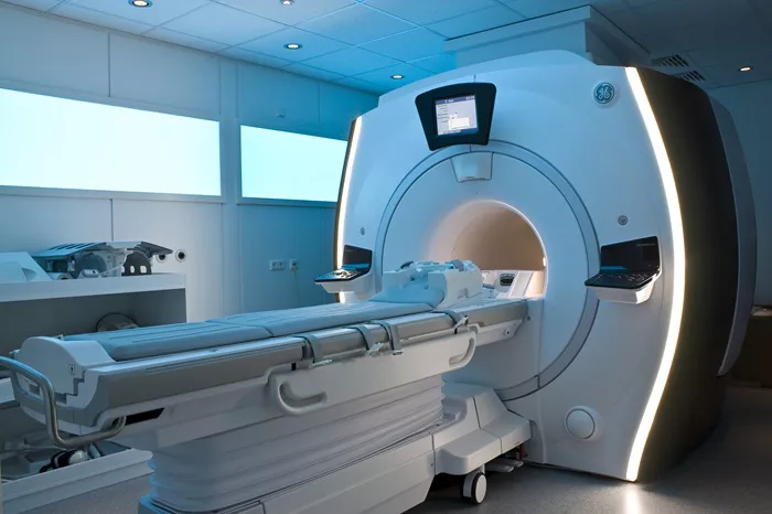
To overcome these challenges, researchers used an advanced magnetic resonance imaging (MRI) technique called ultrahigh-resolution T1 quantitative MRI. This method can capture detailed images of the hypothalamus, revealing small subregions called nuclei.
The Study
The study examined the hypothalamus of 44 young women. Among them, 21 had normal weight, 13 had restrictive anorexia nervosa, and 10 were obese. All participants were matched by age to avoid bias.
Researchers measured the volume and tissue integrity of individual hypothalamic nuclei and analyzed their relation to body mass index (BMI), eating disorder symptoms, hormone levels (ghrelin and leptin), anxiety, and depression.
By combining MRI data with psychological and hormonal measures, the team aimed to uncover how brain changes relate to eating disorders.
Key Findings
The study found distinct differences in the hypothalamic regions of women with anorexia nervosa and obesity compared to those with normal weight. These differences were mainly seen in the para- and periventricular nuclei—areas crucial for regulating feeding behavior—and in the connecting nerve fibers.
Typically, brain volume decreases are linked to aging or disease, but in younger women, larger hypothalamic areas may reflect inflammation or swelling. This swelling might disrupt normal eating signals, increasing the risk of eating disorders.
The study also observed changes in hunger and satiety hormone levels, which correlated with hypothalamic differences and eating disorder severity.
Implications and Future Directions
This new imaging approach helps pinpoint brain changes that may underlie eating disorders in young women. Notably, treatments targeting specific hypothalamic areas, such as glucagon-like peptide 1 (GLP-1) receptor agonists, show promise in improving unhealthy eating patterns.
Researchers suggest that future studies should follow patients over time to see if changes in hypothalamic size or structure happen before symptoms start. Further research into how these brain areas connect with other regions could reveal more about the neural basis of eating disorders.
Related Topics: